Home » What is OA/TOF? » Surgery for OA/TOF
Home » What is OA/TOF? » Surgery for OA/TOF
Surgery to repair OA/TOF is not an emergency, with the rare exception of the ventilated premature baby. It is safer, and better for everyone concerned, to keep a stable OA/TOF baby on a drip overnight and operate during daylight hours the following day.
Correction of OA/TOF involves division of the TOF and then repair of the OA.
This operation is performed in theatre under a general anaesthetic with an experienced paediatric anaesthetist continuously monitoring the baby’s vital signs; heart rate, ECG (the electrical activity in the heart), blood pressure, oxygen saturation (the oxygen-carrying capacity of the blood) and body temperature.
Most babies born with OA undergo a primary repair within the first few days of life. However, approximately 10% of babies have a gap between the upper and lower ends of the oesophagus which is too long for a primary repair. In these cases the surgery will be delayed.
See: Long gap OA – delayed primary anastomosis for more information.
Repair of the oesophageal atresia is usually performed through an opening in the right chest wall called a thoracotomy. The baby is positioned on their side with their arm above the head. The skin is cleaned with antiseptic and an incision made just below the tip of the shoulder blade, following the curve of the ribs around the side of the chest.
The chest cavity is usually entered between the fourth and fifth ribs. The surgeon aims to keep the right lung within its covering membrane (the pleura), carefully sweeping it away from the chest wall.
The first part of the repair involves finding and disconnecting the TOF. Once the fistula is divided, the opening into the trachea is repaired with stitches (sutures).
If the baby is unstable the operation can be stopped safely at this stage and the chest closed to minimise operating time. The thoracotomy can be reopened and the two ends of the oesophagus joined up (anastomosed) another day, when the baby is more stable.
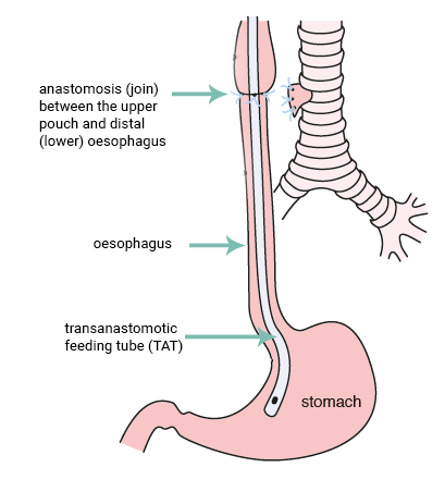
The next step is to identify the upper pouch of the oesophagus. The upper pouch is freed from attachments to the surrounding tissues. The surgeon must assess the gap between the upper and lower ends of the oesophagus to determine if it is possible to join the ends together and repair the atresia by anastomosis. The length of the gap varies.
In most cases, a primary anastomosis (or primary repair of OA) is possible i.e. the two ends can be joined up immediately.
The aim is to repair the oesophagus with as little tension as possible on the ends.
Once most of the sutures are in place, the anaesthetist will usually pass a small feeding tube down the baby’s nose and into the upper oesophagus. The surgeon then guides this tube across the anastomosis into the stomach. This transanastomotic tube or TAT tube allows milk to be fed directly into the stomach during the first few days after the operation, until the baby is well enough to feed by mouth.
After completion of the repair, the chest is closed in layers, with all sutures placed underneath the skin so that no stitches require removal at a later date. Some surgeons leave a chest drain in place for a few days, especially if the repair of the oesophagus was made under tension and there is a concern that the join might leak. In many cases a drain is not required.

Published by TOFS, our leaflets and resources are the must have guides for anyone affected by, or caring for someone with OA/TOF. Order or download your free copies from our shop.
| Cookie | Duration | Description |
|---|---|---|
| cookielawinfo-checkbox-advertisement | 1 year | Set by the GDPR Cookie Consent plugin, this cookie is used to record the user consent for the cookies in the "Advertisement" category . |
| cookielawinfo-checkbox-analytics | 1 year | Set by the GDPR Cookie Consent plugin, this cookie is used to record the user consent for the cookies in the "Analytics" category . |
| cookielawinfo-checkbox-functional | 1 year | The cookie is set by the GDPR Cookie Consent plugin to record the user consent for the cookies in the category "Functional". |
| cookielawinfo-checkbox-necessary | 1 year | Set by the GDPR Cookie Consent plugin, this cookie is used to record the user consent for the cookies in the "Necessary" category . |
| cookielawinfo-checkbox-others | 1 year | Set by the GDPR Cookie Consent plugin, this cookie is used to store the user consent for cookies in the category "Others". |
| cookielawinfo-checkbox-performance | 1 year | Set by the GDPR Cookie Consent plugin, this cookie is used to store the user consent for cookies in the category "Performance". |
| CookieLawInfoConsent | 1 year | Records the default button state of the corresponding category & the status of CCPA. It works only in coordination with the primary cookie. |
| elementor | never | This cookie is used by the website's WordPress theme. It allows the website owner to implement or change the website's content in real-time. |
| enforce_policy | 1 year | PayPal sets this cookie for secure transactions. |
| ts | 3 years | PayPal sets this cookie to enable secure transactions through PayPal. |
| ts_c | 3 years | PayPal sets this cookie to make safe payments through PayPal. |
| Cookie | Duration | Description |
|---|---|---|
| aka_debug | session | Vimeo sets this cookie which is essential for the website to play video functionality. |
| nsid | session | This cookie is set by the provider PayPal to enable the PayPal payment service in the website. |
| player | 1 year | Vimeo uses this cookie to save the user's preferences when playing embedded videos from Vimeo. |
| tsrce | 3 days | PayPal sets this cookie to enable the PayPal payment service in the website. |
| x-pp-s | session | PayPal sets this cookie to process payments on the site. |
| Cookie | Duration | Description |
|---|---|---|
| l7_az | 30 minutes | This cookie is necessary for the PayPal login-function on the website. |
| sync_active | never | This cookie is set by Vimeo and contains data on the visitor's video-content preferences, so that the website remembers parameters such as preferred volume or video quality. |
| Cookie | Duration | Description |
|---|---|---|
| _ga | 2 years | The _ga cookie, installed by Google Analytics, calculates visitor, session and campaign data and also keeps track of site usage for the site's analytics report. The cookie stores information anonymously and assigns a randomly generated number to recognize unique visitors. |
| _gat_UA-51564864-7 | 1 minute | A variation of the _gat cookie set by Google Analytics and Google Tag Manager to allow website owners to track visitor behaviour and measure site performance. The pattern element in the name contains the unique identity number of the account or website it relates to. |
| _gcl_au | 3 months | Provided by Google Tag Manager to experiment advertisement efficiency of websites using their services. |
| _gid | 1 day | Installed by Google Analytics, _gid cookie stores information on how visitors use a website, while also creating an analytics report of the website's performance. Some of the data that are collected include the number of visitors, their source, and the pages they visit anonymously. |
| _hjAbsoluteSessionInProgress | 30 minutes | Hotjar sets this cookie to detect the first pageview session of a user. This is a True/False flag set by the cookie. |
| _hjFirstSeen | 30 minutes | Hotjar sets this cookie to identify a new user’s first session. It stores a true/false value, indicating whether it was the first time Hotjar saw this user. |
| _hjIncludedInPageviewSample | 2 minutes | Hotjar sets this cookie to know whether a user is included in the data sampling defined by the site's pageview limit. |
| _hjIncludedInSessionSample | 2 minutes | Hotjar sets this cookie to know whether a user is included in the data sampling defined by the site's daily session limit. |
| CONSENT | 2 years | YouTube sets this cookie via embedded youtube-videos and registers anonymous statistical data. |
| vuid | 2 years | Vimeo installs this cookie to collect tracking information by setting a unique ID to embed videos to the website. |
| Cookie | Duration | Description |
|---|---|---|
| VISITOR_INFO1_LIVE | 5 months 27 days | A cookie set by YouTube to measure bandwidth that determines whether the user gets the new or old player interface. |
| YSC | session | YSC cookie is set by Youtube and is used to track the views of embedded videos on Youtube pages. |
| yt-remote-connected-devices | never | YouTube sets this cookie to store the video preferences of the user using embedded YouTube video. |
| yt-remote-device-id | never | YouTube sets this cookie to store the video preferences of the user using embedded YouTube video. |
| yt.innertube::nextId | never | This cookie, set by YouTube, registers a unique ID to store data on what videos from YouTube the user has seen. |
| yt.innertube::requests | never | This cookie, set by YouTube, registers a unique ID to store data on what videos from YouTube the user has seen. |
| Cookie | Duration | Description |
|---|---|---|
| _hjSession_2528865 | 30 minutes | No description |
| _hjSessionUser_2528865 | 1 year | No description |
| LANG | 9 hours | No description |